ECG- Rapid Guide
Ischemic Heart Disease
Acute myocardial infarction
ECG-Changes during IAM
An acute, complete obstruction of a coronary artery can lead to a transmural myocardial infarction, also known as a STEMI (ST-segment elevation myocardial infarction). During an acute myocardial infarction (IAM), abnormalities in ventricular depolarization and repolarization can be observed on an electrocardiogram (ECG).
- Initial minutes and within a few hours after the IAM: ST-segment elevatio.
- Next hours: Reduction in the R Wave, and it may begin to appear pathological Q waves (deep and wide Q waves) may begin to appear, reflecting the tissue damage.
- 24 hours later: The ST elevation begins to resolve, and T-wave will become inverted.
- 1 week later: Q wave as pathological and therefore indicative of necrosis, and negative T waves.
- 1 month later: The T wave usually returns to a positive or normal state, indicating improved ventricular repolarization, , in the case of a good evolution of the infarction.
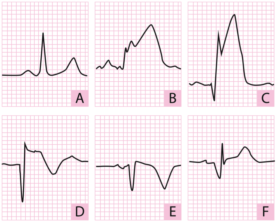 A, normal; B, subepicardial injury wave; C, Q wave indicative of necrosis; D, deeper waves; E, to one week, Q wave persists, and negative T waves; F, in te case of a good outcome, normalized T waves, and Q wave is present.