ECG- Rapid Guide
Introducción
ECG Lead positioning
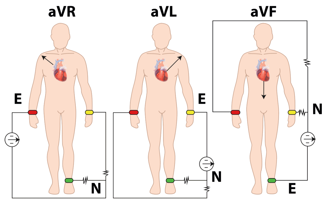
ECG exam typically employs 12 leads across two distinct planes: frontal (DI, DII, DIII, avR, avL, avF) and transverse (V1, V2, V3, V4, V5, V6). Leads may be either bipolar or unipolar. There are 3 bipolar limb leads: I, II and III (bipolar means that the ECG is recorded from two electrodes on the body, that compare electrical potential differences between two electrodes), and 3 augmented unipolar leads: aVR, aVL, aVF (use one electrode as a reference point and a combined reference derived from other electrodes). These leads are obtained using 4 electrodes connected to the ECG machine via 4 wires. The other six leads are considered “precordial leads” because they are placed on the torso (precordium), in the horizontal palne: V1, V2, V3, V4, V5 and V6, that are essentially looking at a specific "slice" of the heart's electrical activity from that electrode's position on the chest. Figures present the placements of ECG leads for correct measurement of the heart's electrical activity. Bipolar leads measure the difference in electrical potential: Lead I, between the electrode of the left arm (exploring electrode) and the electrode of the right arm; Lead II, between the left leg (exploring electrode) and the right arm; Lead III, between the left leg and left arm (wuth the leg electrode being the exploring one).
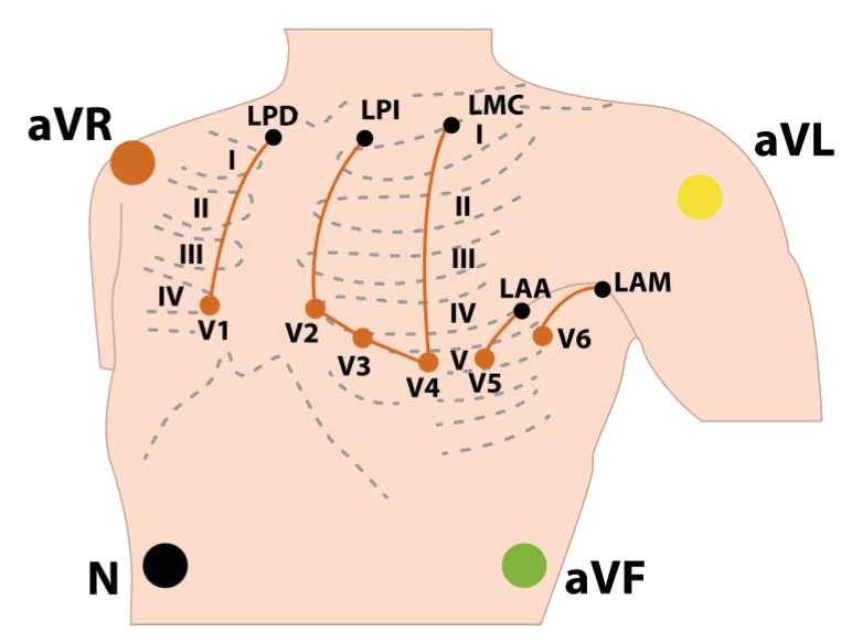
Correct placement for 10 electrodes of a 12-lead ECG (4 for frontal plane, 6 for horizontal plane). The N electrode is designed as neutral electrode, that serves as a grounding point placed on the right leg, minimizing noise and interference during the recording process.